Deciphering the Signals: What Does a Pinched Nerve Look Like on an MRI?
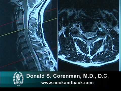
What Does a Pinched Nerve Look Like on an MRI? A pinched nerve, also known as nerve compression or impingement in the medical world, can cause discomfort, pain, and other symptoms.
Magnetic Resonance Imaging (MRI) emerges as a useful tool for visualizing internal structures when seeking a diagnosis.
In this blog post, we’ll look at an MRI to see what a pinched nerve looks like. We seek to provide folks with insights into the diagnostic capabilities of MRI scans and how they reveal telltale indicators of nerve compression by delving into the complexities of medical imaging.
Unveiling the Mysteries of Pinched Nerves
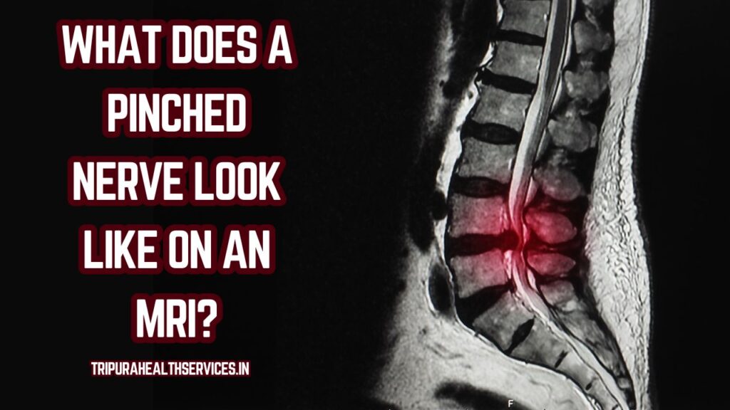
Let’s look at how advanced technology like MRI scans can assist solves the mystery of pinched nerves.
Understanding Pinched Nerves:
A pinched nerve, also known as nerve compression or impingement, happens when a nerve is pressed upon by surrounding tissues such as bones, muscles, or tendons.
Compression can be caused by a variety of reasons, such as herniated discs, bone spurs, or repetitive movements that irritate the nerve. Tingling, numbness, weakness, or pain along the damaged nerve route is common signs of a pinched nerve.
Recognizing the delicate equilibrium within the body’s extensive network of nerves and tissues is necessary for understanding pinched nerves.
These nerves play an important function in signal transmission between the brain and the rest of the body. When excessive pressure disturbs this connection, it can result in a variety of unpleasant and frequently severe symptoms.
Pinched nerves are typically identified using a combination of clinical examination and medical imaging techniques such as Magnetic Resonance Imaging (MRI), which offers precise information about the structures around the afflicted nerves.
What an MRI Reveals About Pinched Nerves:
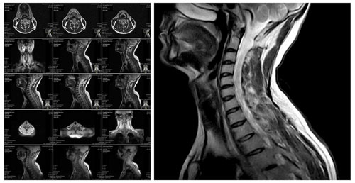
An MRI (Magnetic Resonance Imaging) is a powerful technique for exposing specific details regarding pinched nerves and providing a complete picture of the affected area. An MRI can reveal the following:
- Pinched nerves are frequently caused by structural abnormalities such as herniated discs or bone spurs. An MRI can produce comprehensive images that emphasize these abnormalities, revealing the exact location and amount of the compression.
- Disc Herniation: An MRI can show the displaced disc material and its impact on surrounding nerves if a pinched nerve is caused by disc herniation. Visualizing the size and location of the herniation can assist healthcare practitioners in determining the severity of the problem.
- Nerve Compression: The MRI detects nerve compression by demonstrating a narrowing of the nerve pathway or the presence of neighboring tissues impinging on the nerve. This visual information is critical for comprehending the compression’s specific characteristics.
- Pinched nerves frequently cause inflammation in the affected area. An MRI can identify inflammation, which appears as increased signal intensity around the nerve. This knowledge contributes to a better understanding of the level of tissue irritation and inflammation caused by compression.
- Soft Tissue Detail: MRI excels in producing detailed images of soft tissues, which is critical for diagnosing pinched nerves. It displays the state of the surrounding soft tissues, allowing healthcare experts to assess their role in the compression.
An MRI’s capacity to record both structural and soft tissue information makes it a must-have tool for detecting pinched nerves, assisting healthcare providers in formulating correct treatment strategies customized to the individual characteristics of each case.
Interpreting MRI Findings:
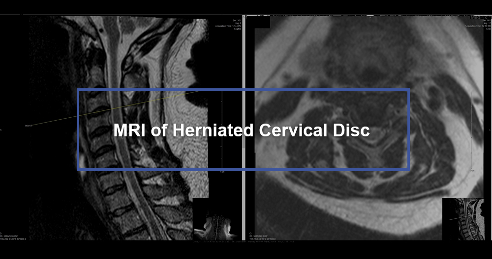
Interpreting MRI findings is a difficult process that necessitates the knowledge of healthcare professionals, notably musculoskeletal or neuroimaging radiologists. Here’s an outline of how MRI findings in the context of pinched nerves are interpreted:
- Compression Level: Radiologists evaluate the degree of nerve compression observed in MRI scans. This entails determining how much surrounding structures, such as herniated discs or bone spurs, are impinging on the nerves.
- Inflammation Present: Pinched nerves are frequently associated with inflammation. Radiologists search for increasing signal intensity near nerves, which indicates inflammation. This information aids in determining the severity of tissue irritation.
- MRI provides a thorough image of the affected area, allowing healthcare experts to examine the impact of pinched nerves on surrounding soft tissues. Muscles, ligaments, and other structures that may be affected by the compression are included.
- Clinical Correlation: Interpreting MRI findings requires more than just visual signals. Clinical correlation entails combining imaging findings with the patient’s symptoms and medical history. This comprehensive approach offers a more accurate diagnosis and assists healthcare providers in developing better treatment regimens.
- Differentiating Between Chronic and Acute Changes: MRI data can help distinguish between chronic and acute changes. Acute nerve compression, such as that induced by trauma, may manifest differently than chronic compression produced by illnesses such as degenerative disc degeneration.
Radiologists and referring physicians must work together to translate MRI findings into relevant insights for patient treatment.
This multidisciplinary approach ensures a comprehensive understanding of the complicated dynamics involved in pinched nerves and guides the creation of tailored treatment options.
FAQs about what does a pinched nerve look like on an MRI?
Q1: Can an MRI definitively diagnose a pinched nerve?
A1: An MRI is a powerful diagnostic technique that can offer detailed images of the tissues around nerves, assisting in the identification of signs of compression.
A definitive diagnosis, on the other hand, frequently necessitates clinical correlation, taking into account symptoms and other diagnostic information.
Q2: What symptoms indicate a pinched nerve?
A2: Pinched nerve symptoms can include tingling, numbness, weakness, or pain along the damaged nerve pathway. The symptoms vary according to the nerve affected and the location of the compression.
Q3: Are there alternatives to an MRI for diagnosing pinched nerves?
A3: While an MRI is a popular and successful imaging tool, other procedures such as nerve conduction studies or electromyography may be utilized to evaluate nerve function and detect areas of compression.
Q4: How long does an MRI for a pinched nerve take?
A4: The length of an MRI for a pinched nerve varies depending on the location being studied. It usually takes between 30 minutes to an hour.
Q5: Is an MRI painful for detecting pinched nerves?
A5: No, an MRI is not painful in and of itself. However, some patients may be bothered by the necessity to remain still during the operation. Communication with the MRI technologist might aid in the management of any issues.
Q6: Can an MRI show the cause of a pinched nerve, such as a herniated disc?
A6: Yes, an MRI is quite good at detecting structural abnormalities such as herniated discs. It can disclose the size, position, and impact of a disc herniation on the nerves around it.
Q7: Can a pinched nerve heal on its own?
A7: A pinched nerve may resolve on its own in some situations with rest and conservative therapy. However, severe or persistent cases may necessitate medical attention.
Q8: How is a pinched nerve treated based on MRI findings?
A8: Treatment is determined by the underlying cause as shown by an MRI. Physical therapy, drugs, lifestyle changes, and, in certain circumstances, surgical intervention are all options for relieving compression.
Q9: Are there risks associated with undergoing an MRI for pinched nerves?
A9: MRI is generally thought to be safe. Individuals with particular medical disorders or metal implants should notify the MRI technologist, and claustrophobia may be taken into account.
Q10: Can an MRI detect pinched nerves in any part of the body?
A10: Yes, an MRI can be used to detect pinched nerves in the spine, extremities, and other sites where nerve compression may occur.
Conclusion:
Finally, an MRI is a useful diagnostic tool for deciphering the complexity of pinched nerves. The pictures generated demonstrate structural anomalies, disc herniation, nerve compression, and evidence of inflammation in the afflicted area.
These visual signals enable healthcare providers to make an accurate diagnosis and correspondingly modify treatment approaches.
As technology advances, the function of MRI in diagnosing and treating pinched nerves becomes increasingly important, providing individuals with a route to comfort and recovery.
We get a better understanding of the junction between medical imaging and the complicated terrain of the human nervous system as a result of this investigation.
- Your Ultimate Guide to Travel Insurance for Adventure Sports
- A Guide to Renters Insurance for Pet Owners: Pet-Proof Your Policy
- Safeguard Your Future: Understanding Identity Theft Insurance
- Safeguard Your Event: Understanding Event Cancellation Insurance
- Everything You Need to Know About Critical Illness Insurance Riders
- Home Equity Loans vs. HELOCs: Which is Right for You?


